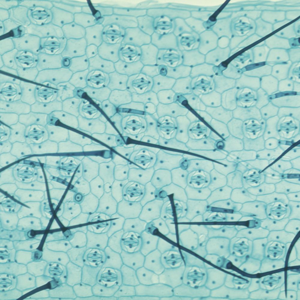
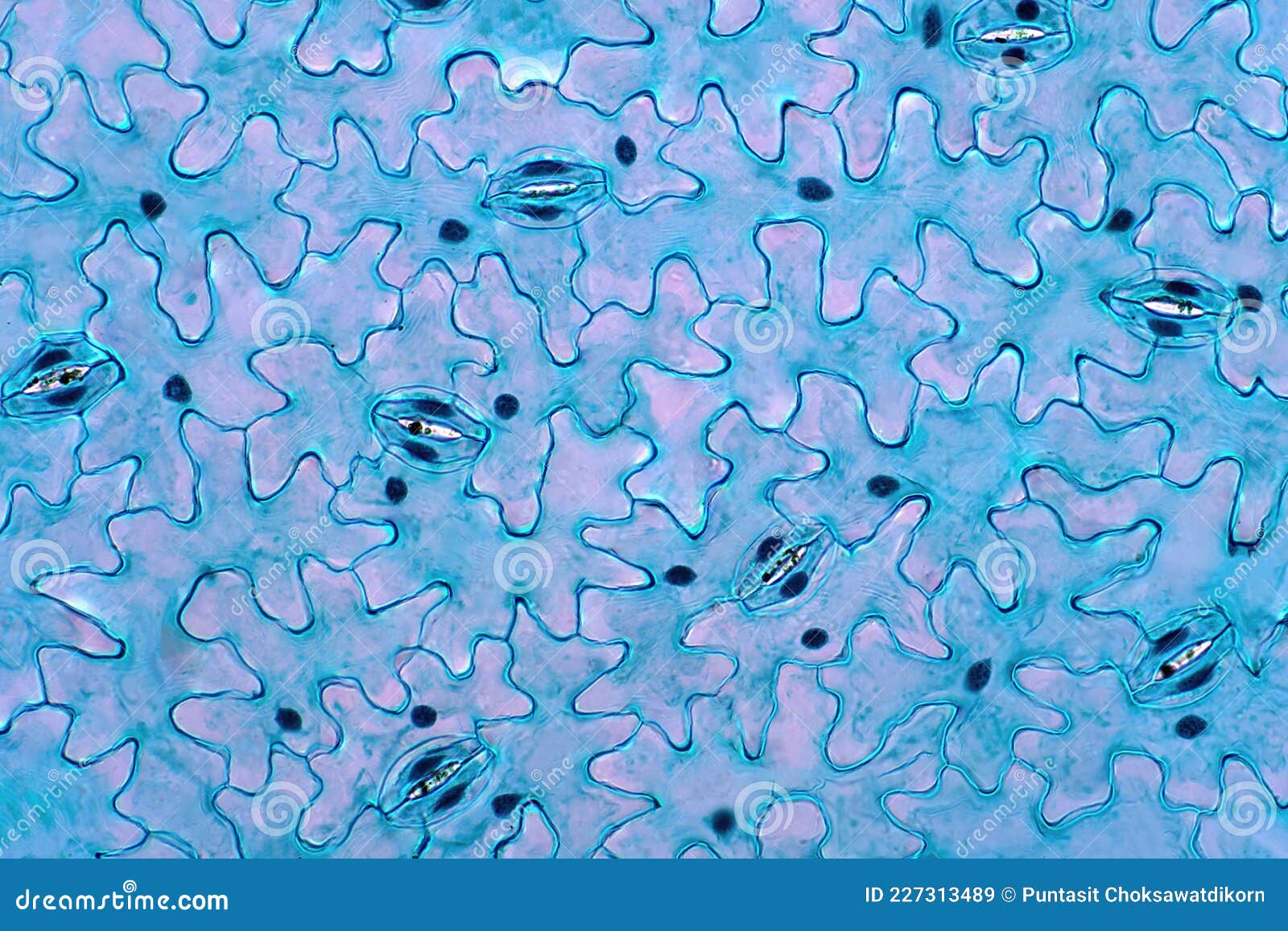
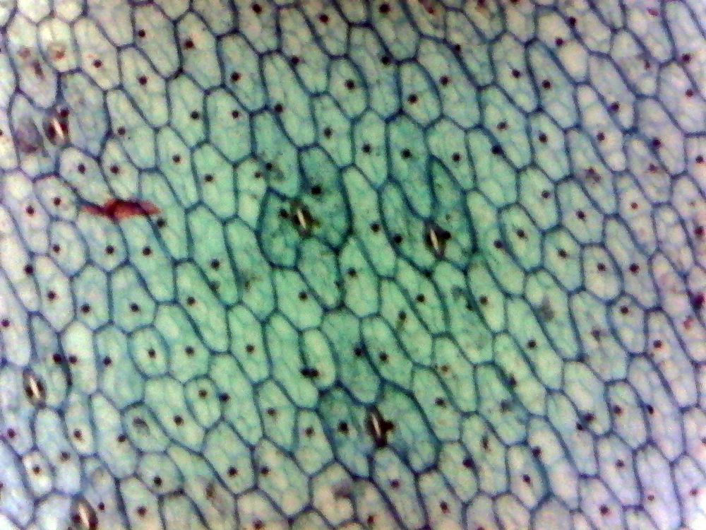
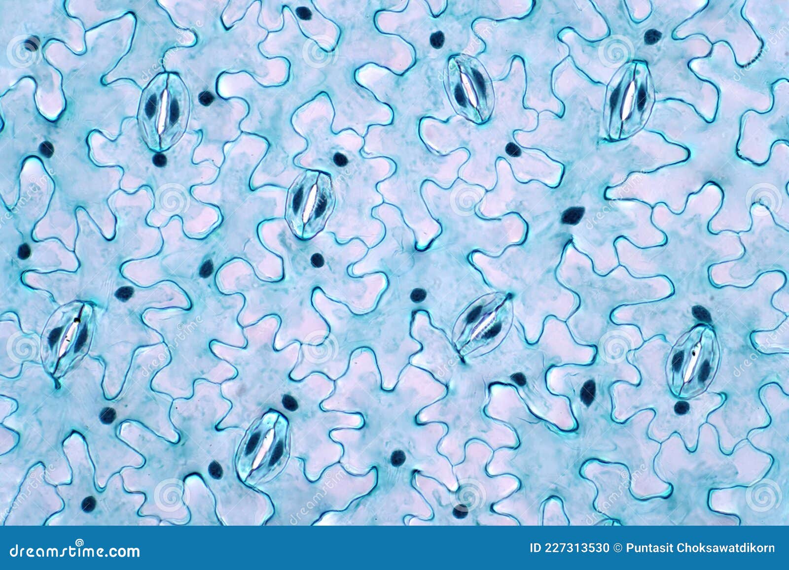
epidermis microscope slide The leaf epidermis under light microscope view has small pores, called
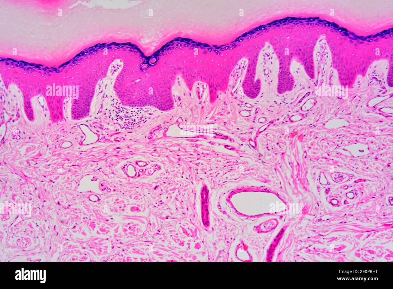
If you are looking for What Does Skin Look Like Under a Microscope? (Images Included) - Optics Mag you’ve came to the right page. We have 35 Pictures about What Does Skin Look Like Under a Microscope? (Images Included) - Optics Mag like Skin epidermis, light micrograph - Stock Image - C039/4787 - Science, Epidermis microscope Stock Vector Images - Alamy and also Micrograph Showing The Epidermis And Dermis Of A Human Finger Skin. The. Read more:
What Does Skin Look Like Under A Microscope? (Images Included) - Optics Mag
 opticsmag.comMonocot Epidermis | Epidermis, Stereo Microscope, Plant Structure
 www.pinterest.commicroscope cell epidermis leaf plant monocot tradescantia slide microscopic stereo structure cells plants theory olympus stomata prepared picture anatomy biologicals
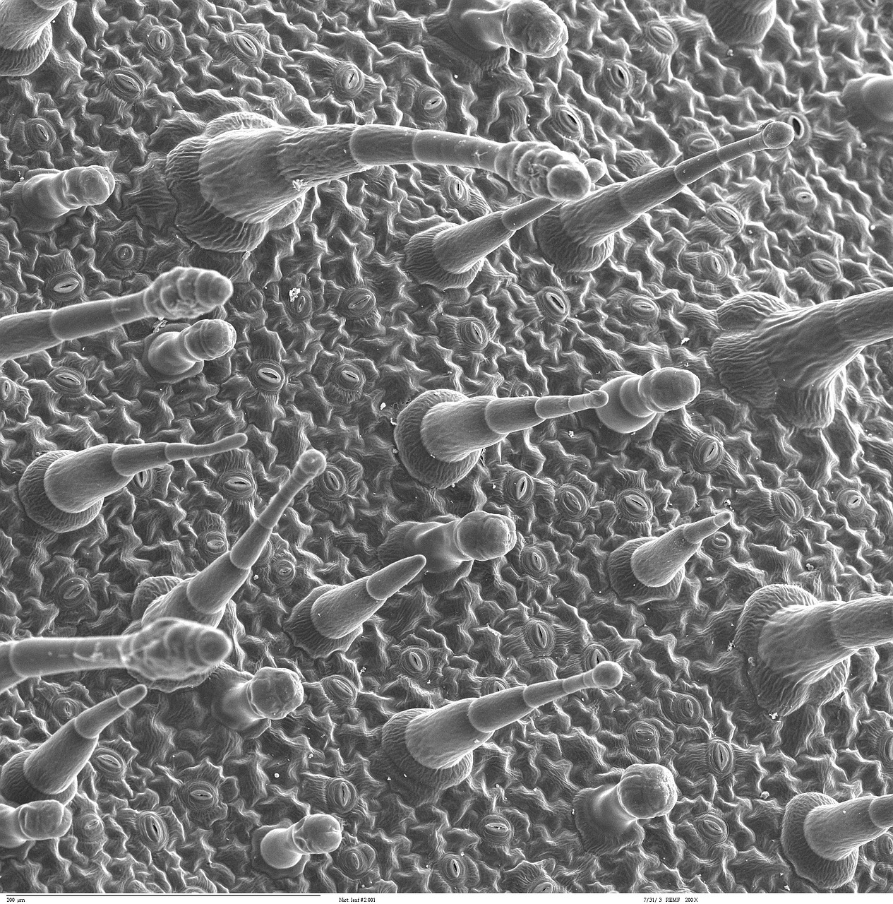
Download Free Photo Of Epidermis,leaf,electron Microscopy,electron
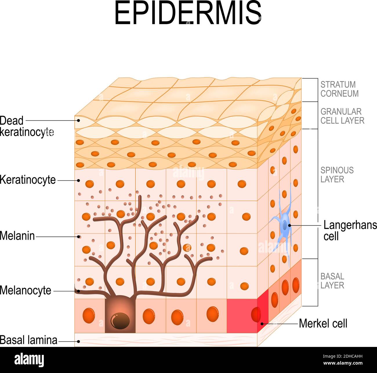
 www.needpix.comA Microscope Image Of The Epidermis. The Epidermis Is Composed Of Four
www.researchgate.netmicroscope epidermis stratum corneum granulosum composed sole spinosum sublayers basale layer lucidum
Layers Of The Skin | Anatomy And Physiology I
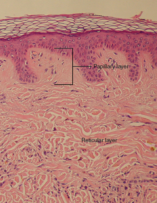 courses.lumenlearning.comskin layers dermis layer papillary cross section slide micrograph reticular
Leaf Epidermis Stomata Under Microscope. Stock Image - Image Of
 www.dreamstime.comepidermis microscope stomata
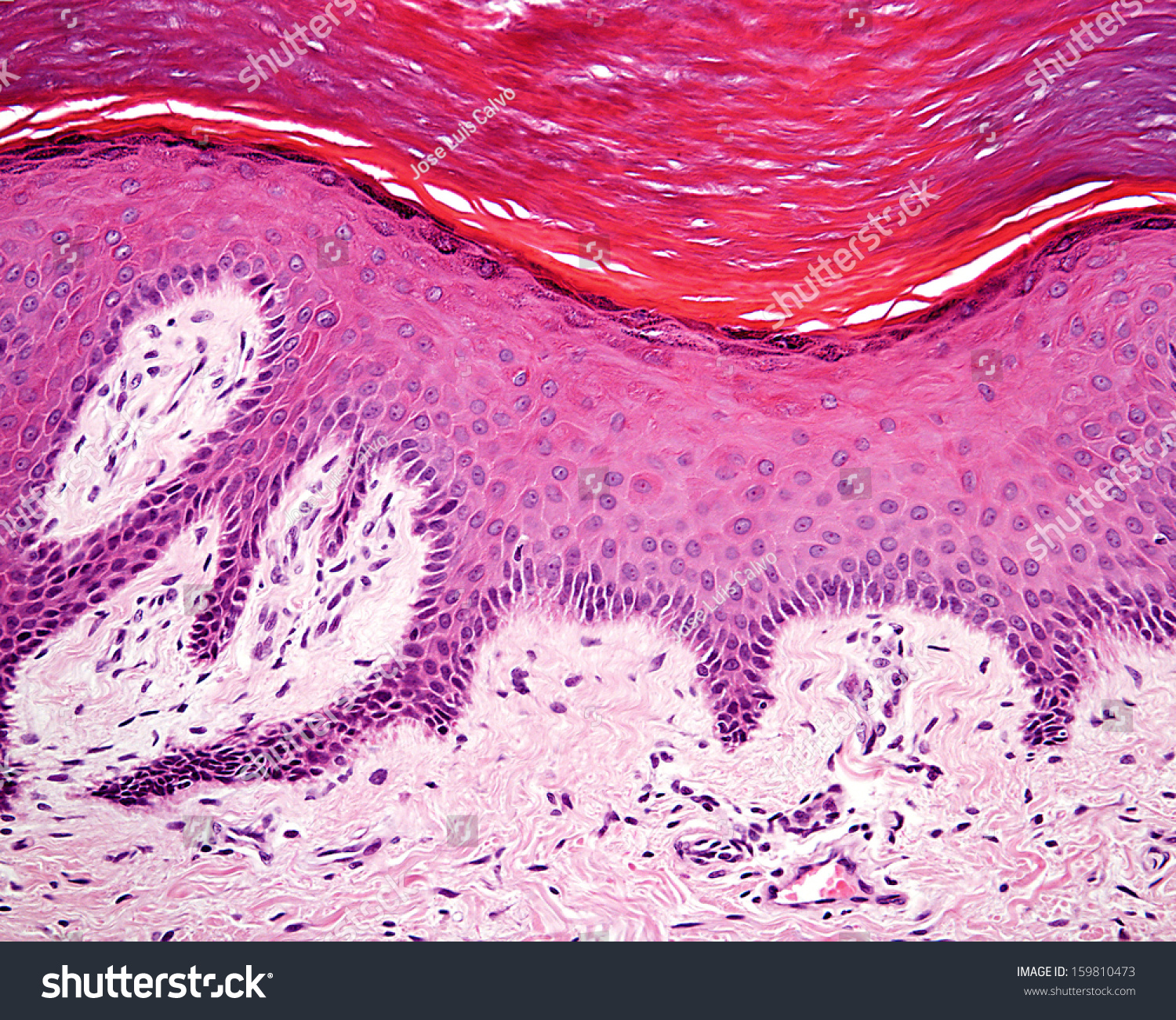
Cross Section Of Human Skin Showing The Stratum Corneum Layer Of The
 www.pinterest.frepidermis microscope stratum corneum microscopic layer gcse cells electron microscopy defences allposters bitesize excretory photographic disease organs excretion
Close Up Plant Epidermis With Stomata Or Leaf Epidermis Under

Layers Of Epidermis (Anonymous, 2020a). | Download Scientific Diagram
 www.researchgate.netSpiderwort Leaf Epidermis, W.m. Microscope Slide | Carolina.com
 www.carolina.comleaf microscope epidermis spiderwort slide tradescantia carolina large
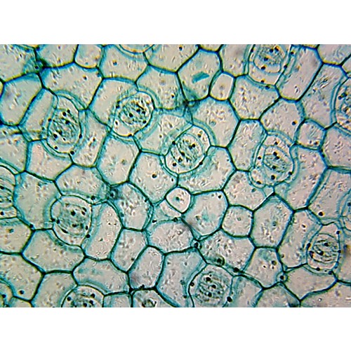
Onion Epidermis, Whole Mount, Cells Of Allium Cepa, 20X Light
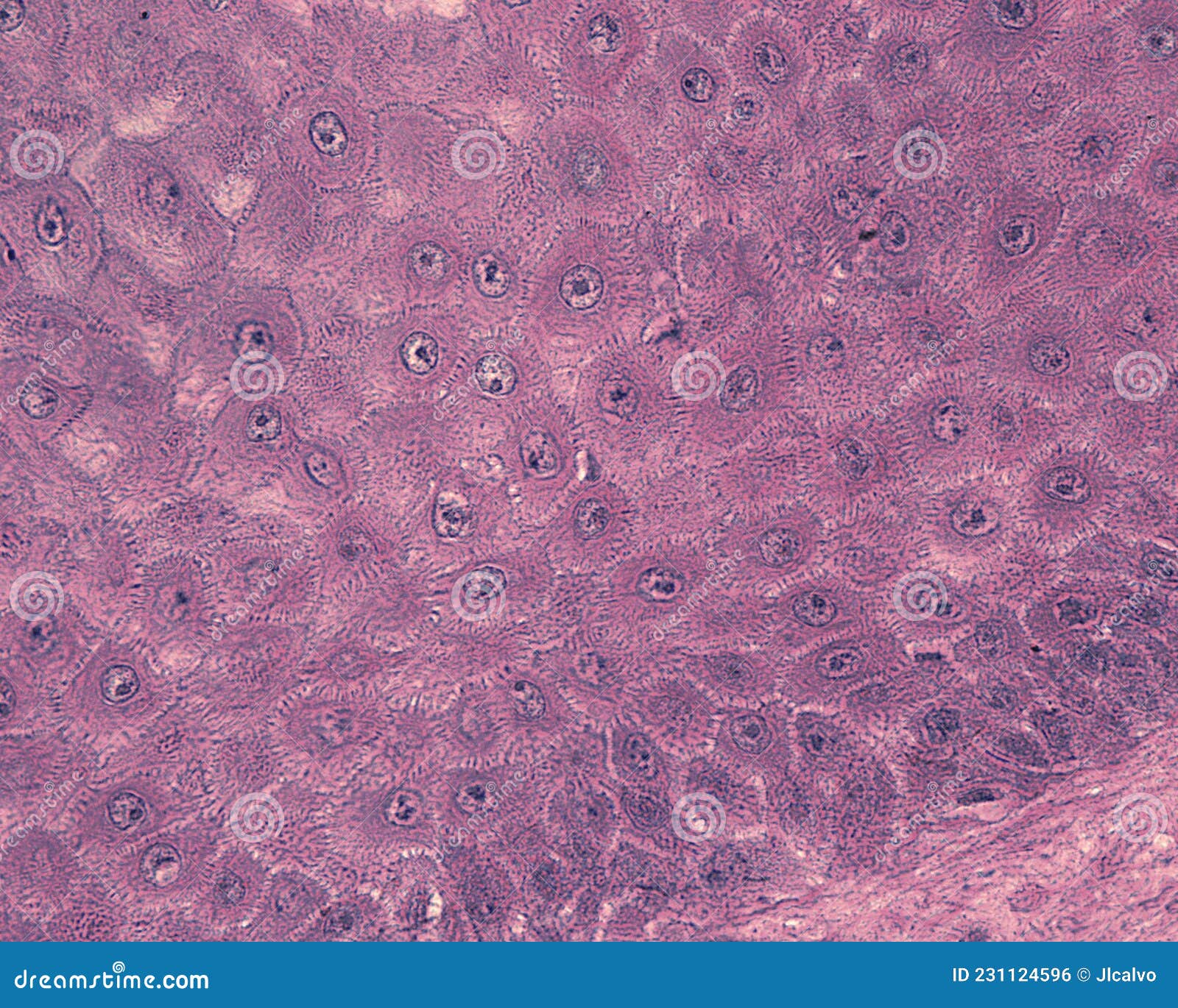
 www.dreamstime.comEpidermis. Spinous Cell Layer Stock Photo - Image Of Cytological
 www.dreamstime.comEpidermal Cells: A Complete Overview – Microscope Clarity
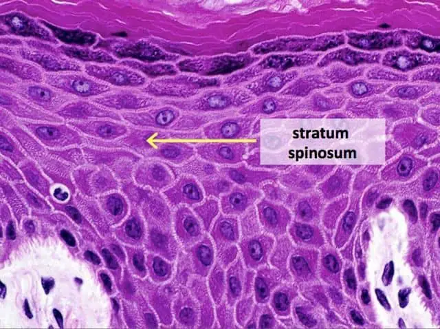 microscopeclarity.comstratum epidermal spinosum microscope labmedicineblog
Dicot Leaf Epidermis, W.m. Microscope Slide | Carolina Biological Supply
 www.carolina.comW.m Microscope Slide Dicot Leaf Epidermis Tillescenter Microscope
 www.tillescenter.orgHuman Skin Epidermis Photograph By Dennis Kunkel Microscopy/science
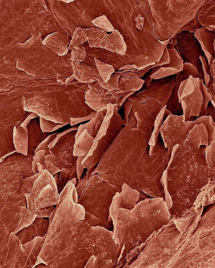 pixels.comskin human epidermis dennis kunkel library microscopy layer science photograph cornified 12th uploaded september which
Staphylococcus Epidermis As Viewed Under A Light Microscope Stock Photo
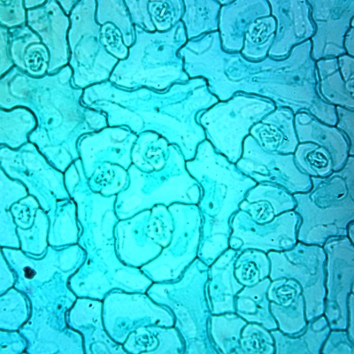
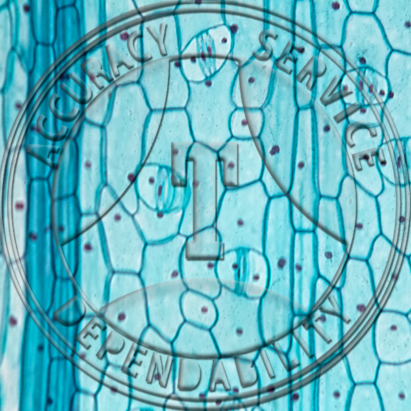
 www.alamy.comOnion Epidermis With Large Cells Under Light Microscope. Clear
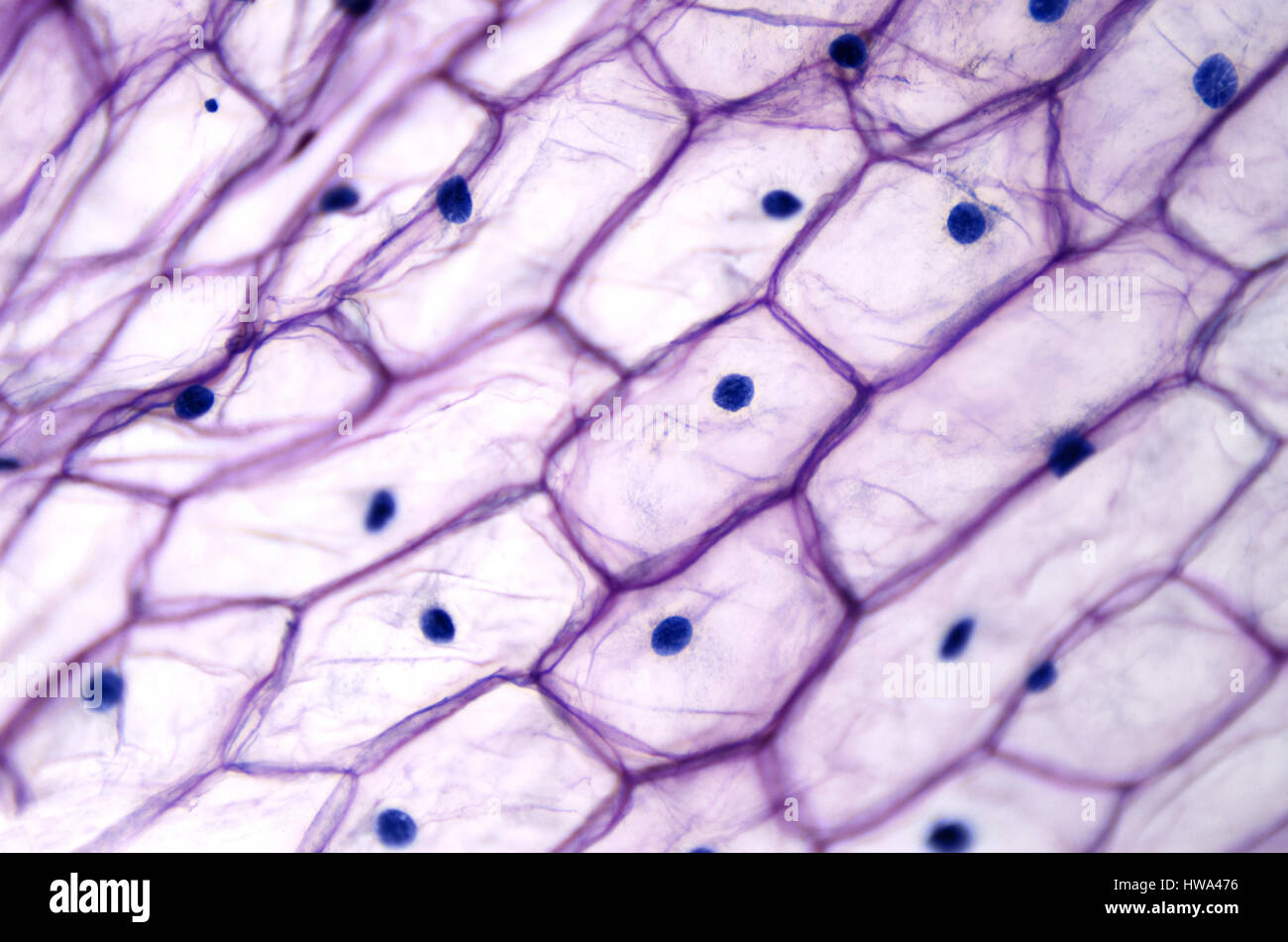 www.alamy.comonion microscope cells under epidermis light epidermal large cell allium cepa vacuole clear layer alamy nucleus
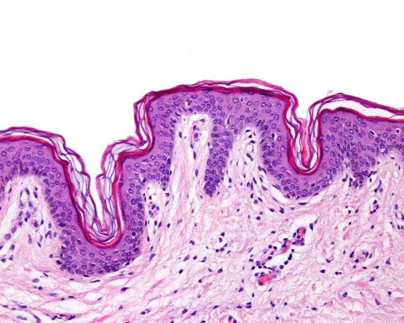
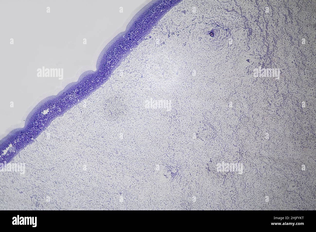
Skin Epidermis, Light Micrograph - Stock Image - C039/4787 - Science
 www.sciencephoto.comepidermis skin micrograph light learn
The Leaf Epidermis Under Light Microscope View Has Small Pores, Called
 www.dreamstime.comThick Skin - Histology | Histology Slides, Nursing Tips, Thick Skin
 www.pinterest.comLeaf Epidermis Prepared Microscope Slide
 www.acornnaturalists.comEpidermis Slide
 ar.inspiredpencil.comEpidermis Microscope Stock Vector Images - Alamy
 www.alamy.comPlant Epidermis Cellulose Cell Walls #2 Photograph By Dennis Kunkel
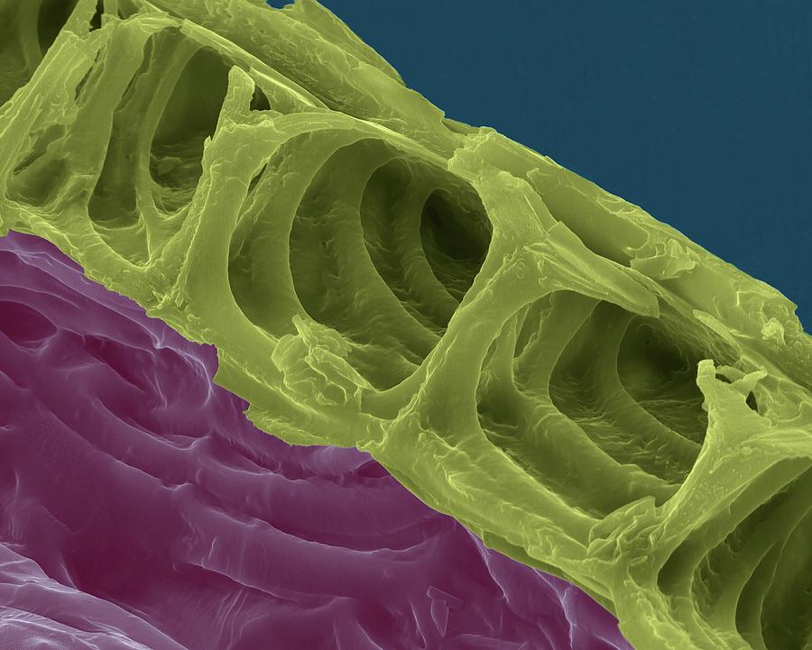 pixels.comcellulose epidermis kunkel dennis microscopy
Micrograph Showing The Epidermis And Dermis Of A Human Finger Skin. The
 www.shutterstock.comepidermis dermis skin layers finger micrograph keratinized squamous human shutterstock epithelium stratified showing search stock
Epidermis Human Skin Hi-res Stock Photography And Images - Alamy
 www.alamy.comskin human epidermis gland sweat stock alamy photomicrograph dermis
W.m Microscope Slide Dicot Leaf Epidermis Tillescenter Microscope
 www.tillescenter.orgHuman Skin - Prepared Microscope Slide - 75x25mm — Eisco Labs
 www.eiscolabs.commicroscope eisco
Onion Bulb (Epidermis, Whole Mount) Prepared Microscope Slide
 www.acornnaturalists.comOnion Bulb, Epidermis - Prepared Microscope Slide | Microscope Slides
 www.pinterest.comW.m Microscope Slide Dicot Leaf Epidermis Tillescenter Microscope
 www.tillescenter.orgPrepared Microscope Slide,Monocot Leaf Epidermis W.M., Showing Stomata
 www.thomassci.comMonocot Leaf Epidermis, W.m., Onion Microscope Slide | Carolina.com
 www.carolina.comleaf epidermis monocot microscope slide onion stomata carolina allium
The Leaf Epidermis Under Light Microscope View Has Small Pores, Called
 www.dreamstime.comOnion microscope cells under epidermis light epidermal large cell allium cepa vacuole clear layer alamy nucleus. Prepared microscope slide,monocot leaf epidermis w.m., showing stomata. Cross section of human skin showing the stratum corneum layer of the